Chapter 20 Biology Class 11 Locomotion and Movement NCERT Solutions
Chapter 20 Biology Class 11 Locomotion and Movement NCERT Solutions
NCERT Textbook Solutions for Class 11 are very important and crusial that helps the students in understanding the complex topics and helps them in the preparation of class 11 board examination as well as verious compititive entrance examinations also. Studying the answers to the questions in the biology textbook will check your understanding of a particular topic and helps you determine your strengths and weaknesses.
Class 11 biology NCERT textbook Solutions Chapter 20 Biology Class 11 Locomotion and Movement NCERT Solutions are provided here with simple step-by-step detailed explanations. Class 11 biology textbook Solutions Chapter 20 Biology Class 11 Locomotion and Movement NCERT Solutions are very popular and come handy for quickly completing your homework and preparing for exams. All questions and answers from the biology textbook Solutions Book of Class 11 biology Chapter 20 are provided here for you for free. You will also love the experience on ybstudy class 11 Solutions. All biology textbook Solutions. Solutions for class 11 These biology textbook solutions are prepared by biology experts and are 100%
Important Points to Remember about Class 11 Locomotion and Movement :
- As we know Movement is common Characteristic feature of all living beings. Protoplasmic streaming, ciliary movements, movements of fins, limbs, wings, etc., are some forms exhibited by animals.
- A voluntary movement which causes the animal to change its place, is called locomotion. Animals move generally in search of food, shelter, mate, breeding ground, better climate or to protect themselves. The cells of the human body exhibit amoeboid, ciliary and muscular movements.
- Locomotion and many other movements require coordinated muscular activities. Three types of muscles are present in our body. Skeletal muscles are attached to skeletal elements. They appear striated and are voluntary in nature.
- Visceral muscles, present in the inner walls of visceral organs are nonstriated and involuntary.
- Cardiac muscles are the muscles of the heart. They are striated, branched and involuntary. Muscles possess excitability, contractility, extensibility and elasticity.
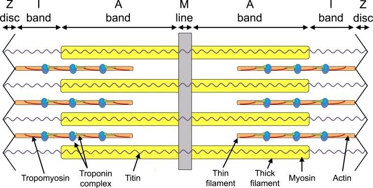
- Muscle fibre is the anatomical unit of muscle. Each muscle fibre has many parallelly arranged myofibrils. Each myofibril contains many serially arranged units called sarcomere which are the functional units. Each sarcomere has a central ‘A’ band made of thick myosin filaments, and two half ‘I’ bands made of thin actin filaments on either side of it marked by ‘Z’ lines.
- Actin and myosin are polymerised proteins with contractility. The active sites for myosin on resting actin filament are masked by a protein-troponin. Myosin head contains ATPase and has ATP binding sites and active sites for actin. A motor neuron carries signal to the muscle fibre which generates an action potential in it. This causes the release of Ca++ from sarcoplasmic reticulum. Ca++ activates actin which binds to the myosin head to form a cross bridge. These cross bridges pull the actin filaments causing them to slide over the myosin filaments and thereby causing contraction. Ca++ are then.returned to sarcoplasmic reticulum which inactivate the actin. Cross bridges are broken and the muscles relax.
- Repeated stimulation of muscles leads to fatigue.
- Muscles are classified as Red and White fibres based primarily on the amount of red coloured myoglobin pigment in them.
- Bones and cartilages constitute our skeletal system. The skeletal system is divisible into axial and appendicular. Skull, vertebral column, ribs and sternum constitute the axial skeleton. Limb bones and girdles form the appendicular skeleton.
- Three types of joints are formed between bones or between bone and cartilage – fibrous, cartilaginous and synovial. Synovial joints allow considerable movements and therefore, play a significant role in locomotion.
Class 11 Locomotion and movement NCERT Solutions :
1. Draw the diagram of a sarcomere of skeletal muscle showing different regions.
Answer:
2. Define sliding filament theory of muscle contraction
Answer: Sliding filament theory which state that contraction of a muscle fibre takes place by the sliding of the thin filaments over the thick filaments.
The sliding filament theory explains how muscles contract. The theory was proposed in 1954, when researchers looked at changes in the sarcomere is the contractile unit of muscle.
There are hundreds of sarcomeres in each muscle fiber and within each sarcomere are contractile proteins known as filaments. There is the thick filament myosin and the thin rope-like filament actin. These filaments slide over the top of the other, causing the muscle to shorten producing a forced contraction.
3. Describe the important steps in muscle contraction.
Answer: The muscular system is responsible for functions such as maintenance of posture, locomotion, and control of various circulatory systems.
The muscular system is closely associated with the skeletal system in facilitating movement. Both voluntary and involuntary muscular system functions are controlled by the nervous system. Muscle contraction describe in following steps
Step 1: Muscle contraction is initiated by signals that travel along the axon and reach the neuromuscular junction or motor end plate. The neuromuscular junction is a junction between a neuron and the sarcolemma of the muscle fibre. As a result, acetylcholine (a neurotransmitter) is released into the synaptic cleft by generating an action potential in the sarcolemma.
Step 2: The generation of this action potential releases calcium ions from the sarcoplasmic reticulum in the sarcoplasm.
Step 3: The increased calcium ions in the sarcoplasm lead to the activation of actin sites. Calcium ions bind to the troponin on actin filaments and remove the tropomyosin, wrapped around actin filaments. Hence, active actin sites are exposed and this allows myosin heads to attach to this site.
Step 4: In this stage, the myosin head attaches to the exposed site of actin and forms cross bridges by utilizing energy from ATP hydrolysis. The actin filaments are pulled. As a result, the H-zone reduces. It is at this stage that the contraction of the muscle occurs.
Step 5: After muscle contraction, the myosin head pulls the actin filament and releases ADP along with inorganic phosphate. ATP molecules bind and detach myosin and the cross bridges are broken.
Step 6: This process of formation and breaking down of cross bridges continues until there is a drop in the stimulus, which causes an increase in calcium. As a result, the concentration of calcium ions decreases, thereby masking the actin filaments and leading to muscle relaxation. The sliding of actin along myosin therefore shortens the sarcomere, causing muscle contraction
4. Write true or false. If false change the statement so that it is true.
(a) Actin is present in thin filament
(b) H-zone of striated muscle fibre represents both thick and thin filaments.
(c) Human skeleton has 206 bones.
(d) There are 11 pairs of ribs in man.
(e) Sternum is present on the ventral side of the body.
Answer :
(a) Actin is present in thin filament – True
(b) H-zone of striated muscle fibre represents both thick and thin filaments – False
Corrected statement: H-zone of the striated muscle fiber represents only thick filaments
(c) Human skeleton has 206 bones – True
(d) There are 11 pairs of ribs in man – False
Corrected statement: There are 12 pairs of ribs in man.
(e) Sternum is present on the ventral side of the body – True
5. Write the difference between:
(a) Actin and Myosin
Answer :
| Actin | Myosin |
|---|---|
| They are thin and short in Size | They are thick and Longer In size |
| Actin filament surface of action Is smooth | Myosin filaments surface of action is hard |
| It is located in cell membrane, Cell wall and Microfilaments. | Myosin is located in Muscles Cells |
| Actin filaments are more abundant | Myosin filaments are less abundant than Actin filament. |
| It carries tropomyosin and troponin | It carry meromyosin |
| Actin filament do not form cross Bridges | Myosin filaments forms cross bridges |
| They are founds in A and I bands | They are found in A bands of sarcomeres |
| They are free at one end | They are free at both ends. |
(b) Red and White muscles
Answer :
| Red Muscle Fibres | White Muscles Fibers |
|---|---|
| In red Muscle fibre more number of Mitochondria are present. | In white Muscles fibre mitochondria is present in less number as compared to red Muscle fibre. |
| In this sorcoplasmic reticulum is less in number. | In white Muscles sorcoplasmic reticulum is present in Large in number. |
| Red Muscles are thin as compared to the white muscle fibre. | White Muscles are thick than white Muscles. |
| In red Muscle fibre the process of contraction is slow as compared to white Muscles. | In white Muscles rate of muscle contraction is fast. |
| Red Muscle fibre use more Oxygen. | It white Muscles fibre less amount of oxygen involved as compared to red Muscle fibre. |
| Red Muscle perform aerobic oxidation without accumulating more lactic acid. | White Muscles fibre perform anaerobic oxidation by accumulating more lactic acid. |
| Example of red Muscle is extensor Muscle. | Example of white Muscles is muscle of the eye. |
| Less amount of energy is produced. | More amount of energy is produced as compared to red Muscle because of presence of glycogen. |
(c) Pectoral and Pelvic girdle
Answer :
| Pectoral Girdle | pelvic Girdle |
|---|---|
| The shoulder girdle or the pectoral girdle is a set of 4 bones which connects to the arm on each side. It is a set of 4 bones -1. 2 scapula2. 2 clavicle | The pelvic girdle consists of two bones: sacrum and the coccyx. The pelvic girdle is formed of paired hip bones each made up of ilium, ischium and pubis. |
| The bones of the pectoral girdle are lighter | It consists of innominate coxal or hip bones |
| It has no articulation with the vertebral column | It has an articulation with the vertebral column. |
| It is also known as shoulder girdle and is found in shoulder on both the sides | It is found in the lower part of the trunk |
| The pectoral girdle is part of the appendicular skeleton which are for the upper limbs. In human beings, the pectoral girdle consists of the scapula and the clavicle. | The pelvic girdle is located in the lower part of the trunk. It is a ring-like bony structure. |
| These are responsible for lifting, holding etc. | These are responsible for jumping, standing etc. |
| It is same in both males and females | Women majorly have gynaecoid pelvis and males have android pelvis |
6. Match Column I with Column II:
| Column I | Column II |
|---|---|
| (a) Smooth Muscle | (i) Myoglobin |
| (b) Tropomyosin | (ii) Thin filament |
| (c) Red Muscle | (iii) Sutures |
| (d) Skull | (iv) Involuntary |
Answer :
| Column I | Column II |
|---|---|
| (a) Smooth Muscle | (iv) Involuntary |
| (b) Tropomyosin | (ii) Thin filament |
| (c) Red Muscle | (i) Myoglobin |
| (d) Skull | (iii) Sutures |
7. What are the different types of movements exhibited by the cells of human body?
Answer : Movement is a characteristic feature of living organisms. Mainly three types of movement exhibited by cells of the human body are:
Amoeboid movement: Amoeboid movement is the most common mode of locomotion in eukaryotic cells. Amoeboid movement is achieved by pseudopodia and involves the flow of cytoplasm as extensions of the organism. Leucocytes present in the blood show amoeboid movement. During tissue damage, these blood cells move from the circulatory system towards the injury site to initiate an immune response. Diverse cell types, including Amoebae, leukocytes, embryonic cells and tumour cells show amoeboid movements.
Ciliary movement: Cilia is the hair-like outgrowth present on the surface of plasma membrane. Ciliary movement generally refers to the rhythmic movement of cilia, which causes movement of the fluid or the cell. Reproductive cells such as sperms and ova show ciliary movement. The passage of ova through the fallopian tube towards the uterus is facilitated by this movement.
Muscular movement: Muscles move body parts by contracting and then relaxing. Muscles can pull bones, but they can’t push them back to the original position. So they work in pairs of flexors and extensors. Muscle cells show muscular movement. The muscular system is an organ system consisting of skeletal, smooth and cardiac muscles. It permits movement of the body,
8. How do you distinguish between a skeletal muscle and a cardiac muscle?
Answer:
Skeletal Muscles :
- Skeletal muscles found in entire bones of the body.
- The length of skeletal muscle is longer than cardiac muscles.
- Gap junction are absent in skeletal muscle.
- The shape of the skeletal muscle is cylindrical.
- skeletal muscles control by somatic nervous system.
- In skeletal muscle cells multiple nucleus is present (multinucleated).
Cardiac Muscles :
- Cardiac muscles found in heart only.
- The length of cardiac muscle is short as compared to skeletal muscle.
- Gap junction present in cardiac muscles.
- Shape of cardiac muscles are semispindle.
- It is controlled by autonomous nervous system.
- In cardiac muscle cells only one nucleus is present (uninucleated).
9. Name the type of joint between the following:-
(a) atlas/axis
(b) carpal/metacarpal of thumb
(c) Between phalanges
(d) femur/acetabulum
(e) Between cranial bones
(f) Between pubic bones in the pelvic girdle
Answer:
(a) atlas/axis – Pivot joint
(b) carpal/metacarpal of thumb – Saddle joint
(c) Between phalanges – Hinge joint
(d) femur/acetabulum – Ball and socket joint
(e) Between cranial bones – Fibrous joint
(f) Between pubic bones in the pelvic girdle – cartilaginous joint
10. Fill in the blank spaces:
(a) All mammals (except a few) have __________ cervical vertebra.
(b) The number of phalanges in each limb of human is __________
(c) Thin filament of myofibril contains 2 ‘F’ actins and two other proteins namely____ and _____.
(d) In a muscle fibre Ca++ is stored in __________.
(e) __________ and __________ pairs of ribs are called floating ribs.
(f) The human cranium is made of __________ bones
Answer :
(a) All mammals (except a few) have seven cervical vertebra.
(b) The number of phalanges in each limb of human is Fourteen
(c) Thin filament of myofibril contains 2 ‘F’ actins and two other proteins namely troponin and tropomyosin
(d) In a muscle fibre Ca++ is stored in Sarcoplasmic reticulum
(e) Eleventh and twelfth pairs of ribs are called floating ribs.
(f) The human cranium is made of eight bones.